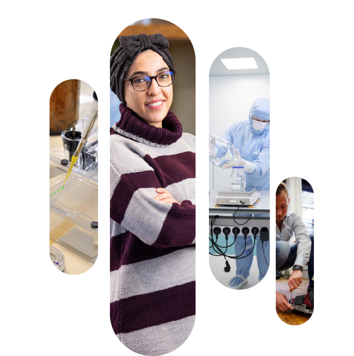
Nawal Aledlbi
4D Local drug distribution determination using advanced optical (light sheet) microscopy
After doing my bachelor’s in applied chemistry, I went for a master’s degree in analytical chemistry. To broaden my horizon, I joined the Nanoscience TopMaster program at the University of Groningen. During my PhD, I look forward to developing a 4D optical analysis to get an insight into targeted drug distribution, that can lead to efficient personalized medicine in IBD patients using light-sheet microscopy.
This project is applied for by Dr. Pieter van der Zaag (FSE) and Prof. Dr. Wouter Nagengast (UMCG).
Project start: September 2022
Unveiling tissue morphology: My journey from chemistry to histology
In the second part of my blog series, I’m excited to explore the challenges of my daily routine. Balancing work between two different groups in distinct locations (Zernike and UMCG) requires constant transitions. At times, I find myself shuttling between these groups up to four times a day—an excellent workout, though not ideal during Groningen’s perpetual rain showers.
On the flip side, transitioning into a completely different field has been a rollercoaster of challenges and discoveries. Each day presents fresh opportunities for learning, and I wouldn’t have it any other way.
Before this journey, here’s a glimpse into my background: I hold a BC in Chemistry and an MC in nanoscience. But histology? Well, It wasn’t even on my radar until this PhD adventure began.
So, why the sudden shift? In my previous blog, I introduced you to the magnificent world of tissue clearing, where chemistry and physics work their magic to render tissues transparent. However, when I presented this groundbreaking science to pathologists and clinicians, their questions came faster than I could blink—questions I couldn’t answer, given my background.
To bridge this knowledge gap, I embarked on a quest to understand tissue morphology. I needed to prove to pathologists that this innovative technology wouldn’t harm tissue structure. Thus, I began my journey into histology, learning how to create paraffinized blocks from tissues before and after becoming transparent. The traditional H&E staining technique, while valuable, couldn’t provide all the answers. Consequently, I delved into various immunohistochemistry staining methods to investigate alterations in different proteins.
As a chemist at heart, I couldn’t help but reminisce about my days in the chemistry lab, crafting molecules and tackling new challenges. In the world of pathology, protocols often provide a clear path to follow. But here’s the twist – I’ve discovered immense joy in this new endeavour when I finally obtain results and embark on the journey of understanding and analyzing them.
Stay tuned for more insights into my PhD adventure. If you’re interested in learning more about my project, feel free to reach out!
Tissue-clearing technology can be like walking a tightrope
As a PhD student studying the drug distribution among inflammatory bowel disease (IBD) patients, I never thought my work would involve making things disappear. However, this is exactly what I’ve been doing lately, using the magic of tissue-clearing technology to make the whole biopsy disappear so we can see what’s going on inside.
Tissue clearing involves using a unique solution, such as (i.e. benzyl Alcohol/benzyl benzoate), to make tissues more transparent, so we can image them more easily by eliminating the light scattering events that happen because of the opaqueness of our tissue which limits the imaging depth. Of course, like any good magic trick, there are some challenges involved, such as, tissue clearing is time-consuming and can be a bit tedious.
Additionally, there are also some technical challenges involved, like making sure the tissue structure, as well as the fluorescent dyes used to label the drugs, are not affected by the clearing process.
It’s kind of like walking a tightrope – we have to be careful not to disrupt the delicate balance of the tissues. But despite these challenges, I’ve actually had much fun working with tissue-clearing technology. It’s like being a magician, only instead of pulling rabbits out of hats, I’m making biopsies disappear.
And the best part is: This work could actually help improve treatment for IBD patients.
By using tissue-clearing technology, we can develop a better understanding of how drugs are distributed in the gastrointestinal tract. This, in turn, can lead to personalized treatment plans that are tailored to each patient’s unique needs. It’s exciting to be part of a project that could have such a positive impact on people’s lives.
So that has been quite a challenging and exciting ride, from working with pig’s colon to developing technology for personalized treatment.
If you’re interested in learning more about my adventures in tissue clearing, feel free to reach out – I’m always happy to chat about the magic of science!